

KPV is a tripeptide with the sequence Lys-Pro-Val (lysine-proline-valine) corresponding to the C-terminal fragment of the α-MSH hormone at positions 11–13. α-MSH is a thirteen-amino-acid melanocortin peptide classically associated with skin pigmentation, but is also a potent endogenous anti-inflammatory mediator. Studies have shown that the anti-inflammatory and antimicrobial effects of the hormone are concentrated in its short C-terminal fragment – the KPV tripeptide. This allows us to separate the immunomodulatory effect from the pigmentation effect, which cannot be separated in full α-MSH.
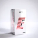
KPV
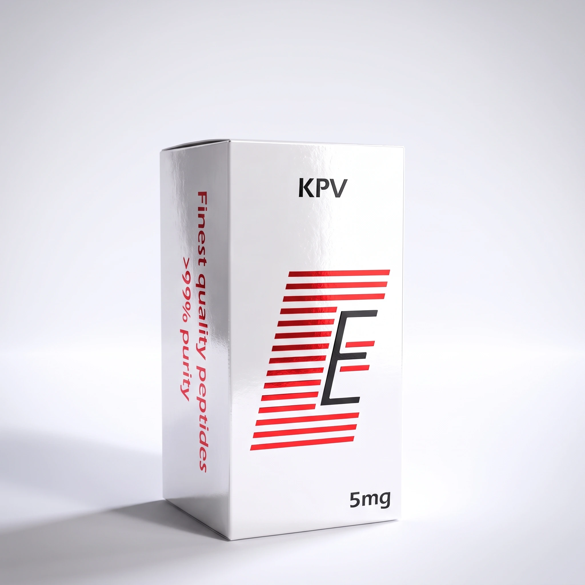
KPV (Lys-Pro-Val) — anti-inflammatory tripeptide, C-terminal fragment of α-MSH (11–13), for laboratory tests. Described in the literature as an intracellular inhibitor of the NF-κB pathway (reduction of IL-1, TNF-α, IL-6) and a substrate of the PepT1 transporter, particularly explored in models of intestinal inflammation. RUO reagent
25,99 €
Fast shipping
Delivery options
Returns policy
KPV (Lys-Pro-Val) - anti-inflammatory tripeptide - RUO research reagent
- KPV (Lys-Pro-Val) — anti-inflammatory tripeptide, C-terminal fragment of α-MSH (11–13), for laboratory tests.
- Described in the literature as an intracellular inhibitor of the NF-κB pathway (reduction of IL-1, TNF-α, IL-6) and a substrate of the PepT1 transporter, particularly explored in models of intestinal inflammation.
KPV (Lys-Pro-Val) is not currently on the WADA Prohibited List as a separate substance mentioned by name. This does not relieve the researcher from verifying current guidelines – anti-doping lists are updated annually, and collective categories (e.g. S0 – unapproved substances) may include compounds without mentioning them by name. Registered athletes (ADAMS) must check the current list of prohibited substances before making any decision regarding use. In the Pro-Body catalog KPV appears only as a RUO research reagent, not as an adjuvant.
KPV from the Endogenic line is anti-inflammatory tripeptide — the shortest active fragment of the hormone α-melanotropin (α-MSH), composed of three amino acid residues: lysine, proline and valine. Unlike most regenerative peptides, KPV is not designed for the reconstruction of connective tissue, but for one, well-defined direction of research: silencing the inflammatory response at the cell level.
This makes it an unusual molecule in the catalog of reagents – short, stable and molecularly precise. The entire history of KPV begins with a question that has been puzzling researchers for years: is it possible to separate the anti-inflammatory effect of the α-MSH hormone from its classic effect on melanocortin receptors – responsible, among other things, for skin pigmentation.
The answer turned out to be the KPV tripeptide, i.e. the C-terminal fragment of α-MSH (positions 11–13). It has been described in the preclinical literature as a compound that retains a significant portion of the anti-inflammatory and antimicrobial activity of the parent hormone while losing its effect on pigmentation. In other words: the inflammation-silencing effect without the tanning effect.
This distinction has practical implications for experimental design. KPV is particularly explored in models in the literature intestinal inflammation (colitis, models of inflammatory bowel disease – IBD), where the PepT1 transporter present in the intestinal epithelium plays a central role. This format is provided as vial of lyophilisate — sterile, dry peptide for self-reconstitution in laboratory conditions, giving the researcher full freedom to select the working concentration for a specific experimental system.
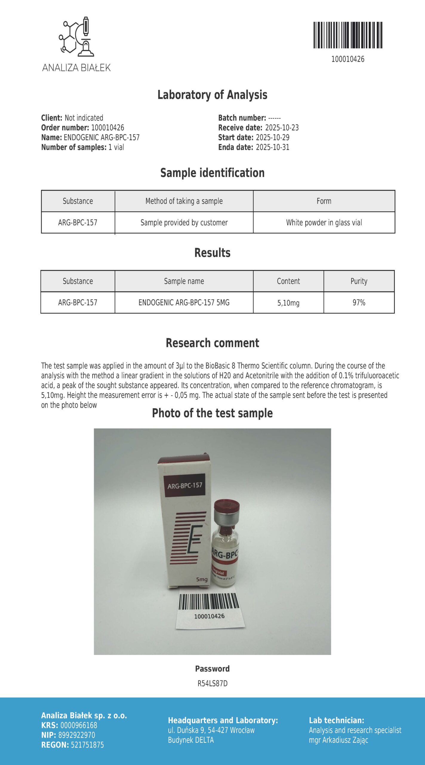
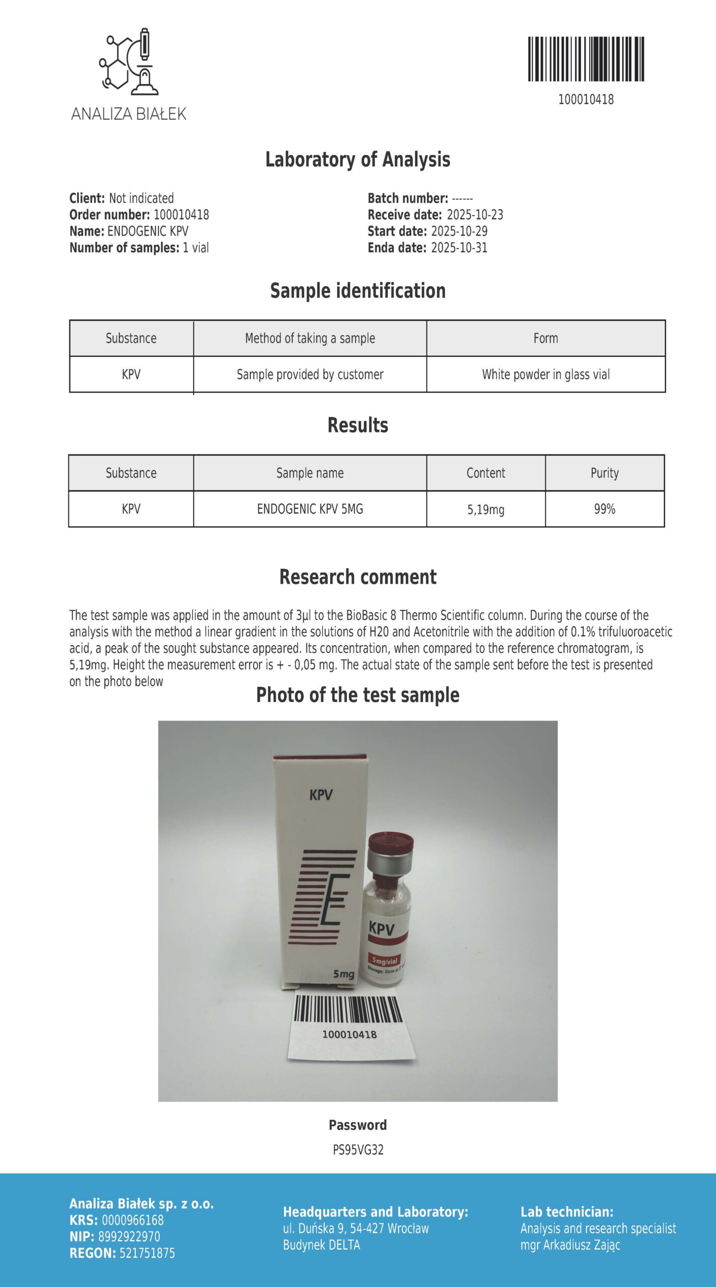
Purity verified by HPLC ≥98%, identity confirmed by Q-TOF mass spectrometry, COA available for each batch. The Endogenic KPV line also appears in other forms (including capsules in research blends and topical preparations). This description applies basic form – vials of lyophilisate as RUO reagent – and this is the subject of the following characterization.
General description - α-MSH and its anti-inflammatory fragments
α-MSH (melanotropic hormone, alpha-melanotropin) is a thirteen-amino-acid peptide belonging to the family melanocortin — derivatives of the precursor proopiomelanocortin (POMC). It is classically associated with skin pigmentation because it activates the melanocortin receptor MC1R in melanocytes. However, α-MSH has a much broader function in the body: it is one of the strongest endogenous anti-inflammatory mediators, described in the literature as a compound that inhibits the production of pro-inflammatory cytokines in many types of cells.
A breakthrough in this context was the observation that the anti-inflammatory effect of α-MSH is concentrated in its C-terminal fragment — Lys-Pro-Val tripeptide (KPV, items 11–13). This fragment retains anti-inflammatory and antimicrobial activity, but – importantly – it no longer shows affinity for the receptors responsible for pigmentation in a way comparable to the full hormone.
This allowed for the research separation of the immunomodulatory effect from the pigmentation effect, which cannot be separated in full α-MSH. Anti-inflammatory peptides constitute a distinct class of molecular tools. Unlike regenerative peptides (such as BPC-157 or TB-500), whose main described mechanism is angiogenesis and matrix reconstruction, KPV-type melanocortin peptides are explored in the literature primarily as modulators NF-κB pathway — central switch of inflammatory gene expression.
In the Pro-Body catalog, KPV is among the research tools for immunomodulation and the intestinal axis – an area that also includes other peptides for immunity, complementary to Pillar I (regenerative peptides).
What is KPV - Lys-Pro-Val tripeptide, α-MSH fragment
KPV it tripeptide with the sequence Lys-Pro-Val (lysine-proline-valine; single-letter notation KPV), corresponding to the C-terminal fragment of α-MSH at positions 11–13. It is one of the shortest active molecules in the class of melanocortin peptides – just three amino acid residues, yet retaining the measurable biological activity of the parent hormone. The short chain length has practical consequences.
The tripeptide is a relatively small and lipophilic molecule, which has been associated in the literature with its ability to penetration into the cell and intracellular action – and not only through surface receptors. The proline residue in the central position gives the molecule characteristic conformational rigidity, typical of short peptides with a proline motif, which may affect its stability and molecular interactions.
In the literature, KPV has been described in several complementary contexts: as a direct inhibitor of the NF-κB pathway inside the cell, as a substrate of the PepT1 transporter in the intestinal epithelium (which makes it interesting in models of intestinal inflammation), and as a fragment retaining the antimicrobial activity characteristic of melanocortin peptides.
Chemical characteristics
| Parameter | Value |
|---|---|
| Name | KPV (Lys-Pro-Val), C-terminal fragment of α-MSH (11–13) |
| Line | Endogenous |
| Class | Anti-inflammatory tripeptide, melanocortin derivative |
| Sequence (1 letter) | KPV |
| Sequence (3-letter) | Lys-Pro-Val (lysine-proline-valine) |
| Number of amino acids | 3 |
| Summary formula | C₁₆H₃₀N₄O₄ [to be verified by COA] |
| Molar mass | ~342.4 g/mol [to be verified in COA] |
| CAS number | 67727-97-3 |
| Origin of the sequence | C-terminus of α-MSH (positions 11–13) |
| Physical form | Lyophilisate (sterile powder for reconstitution) |
| HPLC purity | ≥98% |
| Identity confirmation | Q-TOF mass spectrometry |
Mechanism of action at the molecular level
In the preclinical literature, KPV is described as a compound with a multidirectional but consistently anti-inflammatory profile. The literature indicates five complementary axes of molecular activity. Below, each with the name of the route and the practical ratio observed in the research models.
Anti-inflammatory effect independent of melanocortin receptors
This is the central axis of the KPV profile. Unlike full α-MSH, some of whose effects occur through melanocortin receptors on the cell surface, KPV has been described as a compound penetrating into the cell and acting intracellularly. W badaniach na liniach komórkowych (Luger i wsp. 2007; Brzoska i wsp. 2008) KPV wiązano z hamowaniem szlaku NF-κB and signaling MAPK — two main cascades triggering the expression of inflammatory genes.
The effect observed in the models was a reduction of pro-inflammatory cytokines: IL-1, TNF-α and IL-6. In experimental practice, this meant silencing the inflammatory response without the need to activate pigment receptors.
Transport across the membrane and the PepT1 transporter – intestinal axis
The second axis explains the particular interest in KPV in models of intestinal inflammation. PepT1 is a di- and tripeptide transporter present in the membrane of intestinal epithelial cells (enterocytes). In intestinal inflammation (colitis, IBD models), PepT1 expression changes, and this transporter can actively transport KPV into epithelial cells. Dalmasso et al. (2008, Gastroenterology) described KPV as a PepT1 substrate, the transport of which into enterocytes was associated with local anti-inflammatory effects in the intestinal mucosa.
This makes the tripeptide a particularly interesting tool for research on the intestinal axis – KPV can reach the interior of epithelial cells where the inflammatory process takes place.
Inhibition of NF-κB nuclear activation and translocation
The third axis details the mechanism of the first. NF-κB in an inactive cell remains in the cytoplasm, bound to the inhibitor. The inflammatory signal releases NF-κB, which moves to the cell nucleus (nuclear translocation) and there it triggers the transcription of inflammatory genes. In studies, KPV has been described as a compound that inhibits this translocation – keeping NF-κB out of the nucleus and thus limiting the initiation of the inflammatory cascade at the source. This is an intracellular capture point, consistent with the reported ability of the tripeptide to enter the cell.
Antimicrobial effect
The fourth axis concerns antimicrobial activity. Melanocortin peptides, including α-MSH fragments, retain some antimicrobial activity in the literature – Cutuli et al. (2000) described KPV in the context of inhibiting the growth of selected microorganisms (bacteria and fungi) in in vitro models. KPV, as the shortest active fragment, retains this feature of the parent hormone, which makes it interesting in research on peptides with a dual profile: anti-inflammatory and antimicrobial at the same time.
Support the reduction of tissue inflammation in models
The fifth axis is a wider, tissue translation of the previous four. In inflammatory models (skin, intestine, connective tissue), the reduction of the NF-κB signal and the reduction of pro-inflammatory cytokines were associated with the reduction of local inflammation and – indirectly – with a better course of reparation in the inflamed area (Kannengiesser et al. 2008).
This is an effect observed at the tissue level as a result of molecular mechanisms, not a separate pathway. Reference data mainly come from work on melanocortins and their fragments (Luger et al. 2007; Brzoska et al. 2008) and from studies on the intestinal axis and the PepT1 transporter (Dalmasso et al. 2008; Kannengiesser et al. 2008).
IMPORTANT DISTINCTION
“Anti-inflammatory” or “silences intestinal inflammation” in the context of this description means: “in vitro and in vivo studies in animal models have observed the effect of KPV on the NF-κB pathway, MAPK signaling and on markers of pro-inflammatory cytokines (IL-1, TNF-α, IL-6), and in intestinal models – transport via PepT1.” This is not a guarantee of effect in humans or a suggestion that the KPV reagent may act as a treatment for inflammation, intestinal diseases or infections. All described mechanisms refer to preclinical models – the reagent remains a molecular tool for laboratory research.
Important distinction - KPV, melanocortins and regenerative peptides
KPV (anti-inflammatory effect without activation of MC receptors) vs MT-II and PT-141 (melanocortin agonists). KPV is a fragment of α-MSH, but – unlike full melanocortin agonists – it has been described as an anti-inflammatory compound acting mainly intracellularly, without classic activation of melanocortin receptors (MC1R–MC5R) responsible for pigmentation or stimulation. This distinguishes it fundamentally from a peptide Melanotan 2, a pan-agonist of MC receptors responsible for pigmentation, and from PT-141 (bremelanotide), an MC4R agonist.
KPV retains the anti-inflammatory and antimicrobial fragment of the parent hormone, but does not cause a pigmentation effect – which in studies allows the separation of immunomodulation from receptor action.
KPV vs BPC-157 (different mechanisms, synergistic research stack). BPC-157 is a regenerative pentadecapeptide whose main described mechanism is angiogenesis (VEGF/VEGFR2) and reconstruction of connective tissue – in the catalog it corresponds to injectable BPC-157 10mg. KPV works differently: its target is the NF-κB pathway and silencing pro-inflammatory cytokines. In the context of the intestinal axis, both peptides are sometimes considered in the literature as complementary research tools — BPC-157 as a cytoprotective and pro-angiogenic compound, KPV as a direct modulator of inflammation.
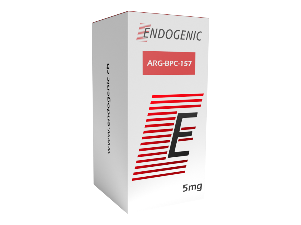
This is why research blends combining both peptides, such as: ARG-BPC-157 + KPV in the form of capsules. The mechanisms are different, but research-synergetic – they do not replace each other, but complement each other.
Salts and forms. KPV is available on the market in various forms (free powder, salts, forms for top application). The core sequence (Lys-Pro-Val) remains identical, but the salt form and product form change the physicochemical parameters and solubility. This product is vial of lyophilisate — sterile reconstitution powder, the identity and mass of which shall be determined by the COA of the batch.
Applications in scientific research
KPV as an RUO reagent is used in several research directions focused on immunomodulation and the intestinal axis. The form of a lyophilized vial works especially well where the researcher needs complete freedom in selecting the working concentration for a specific experimental system.
Models of intestinal inflammation (colitis, IBD) and the PepT1 axis. Models of DSS or TNBS-induced colitis in rodents, cultures of intestinal epithelial cell lines (Caco-2, HT-29). Endpoints: mucosal inflammatory index, level of proinflammatory cytokines (IL-1, TNF-α, IL-6), expression and activity of the PepT1 transporter, NF-κB activation. This is historically the most explored area of KPV research – Dalmasso et al. (2008) and Kannengiesser et al. (2008) described the tripeptide in this context.
The NF-κB pathway and inflammatory signaling. Cultures of immunocompetent cells (macrophages, monocytes) and keratinocytes. Endpoints: NF-κB nuclear translocation, MAPK kinase phosphorylation, cytokine profile. KPV functions as a reference inhibitor of the NF-κB pathway in comparative work on anti-inflammatory compounds.
Antimicrobial activity of melanocortin peptides. In vitro models of bacterial and fungal growth inhibition (Cutuli et al. 2000). Endpoints: minimum inhibitory concentration (MIC), microbial growth curves. KPV, as the shortest active fragment of α-MSH, retains this activity, which makes it a model for research on dual-profile peptides.
Comparative studies in the class of melanocortins. KPV as a reference compound when testing the separation of anti-inflammatory activity from receptor activity (pigmentation, stimulation) within the melanocortin family – in combination with full α-MSH and MC agonists.
Summary
KPV (Lys-Pro-Val) is an anti-inflammatory tripeptide – the C-terminal fragment of the α-MSH hormone at positions 11–13, composed of lysine, proline and valine (CAS 67727-97-3; C₁₆H₃₀N₄O₄; ~342.4 g/mol – values to be confirmed in the COA). It is supplied in the form of a lyophilized vial for self-reconstitution in laboratory conditions. Five axes of tripeptide molecular activity have been described in the preclinical literature: anti-inflammatory activity independent of melanocortin receptors (inhibition of NF-κB and MAPK, reduction of IL-1, TNF-α, IL-6), transport by PepT1 in the intestinal epithelium important in models of intestinal inflammation, inhibition of NF-κB nuclear translocation, antimicrobial activity and reduction of tissue inflammation in models.
What makes KPV an unusual molecule is the molecular separation of the anti-inflammatory effect from the receptor action of the parent hormone – the tripeptide retains the immunomodulatory and antimicrobial effect, but does not cause the pigmentation characteristic of melanocortin agonists such as Melanotan 2. In the context of the intestinal axis, KPV is sometimes considered in the literature as a complementary tool to BPC-157 – a different mechanism, but research is synergistic.
The regulatory profile remains clear: KPV is not registered as a medicine (no EMA/FDA/EFSA authorization). Pro-Body is listed in the Pro-Body catalog as a Research Use Only reagent – with HPLC purity ≥98%, MS Q-TOF confirmation and COA for each batch. The broader context of peptide immunomodulation and the selection of molecules supporting the immune system is discussed in the review peptides for immunity.
Bibliography
- Luger TA, Brzoska T (2007). alpha-MSH related peptides: a new class of anti-inflammatory and immunomodulating drugs. PubMed
- Dalmasso G, Charrier-Hisamuddin L, Nguyen HT, Yan Y, Sitaraman S, Merlin D (2008). PepT1-mediated tripeptide KPV uptake reduces intestinal inflammation. PubMed
- Kannengiesser K, Maaser C, Heidemann J, Luegering A, Ross M, Brzoska T, et al. (2008). Melanocortin-derived tripeptide KPV has anti-inflammatory potential in murine models of inflammatory bowel disease. PubMed
- Brzoska T, Luger TA, Maaser C, Abels C, Böhm M (2008). Alpha-melanocyte-stimulating hormone and related tripeptides: biochemistry, antiinflammatory and protective effects in vitro and in vivo, and future perspectives for the treatment of immune-mediated inflammatory diseases. PubMed
- Cutuli M, Cristiani S, Lipton JM, Catania A (2000). Antimicrobial effects of alpha-MSH peptides. PubMed
Chemical reagent Research Use Only. KPV (Lys-Pro-Val, lyophilisate vial, Endogenic line) in the Pro-Body catalog is not a medicinal product, dietary supplement or food. It is not intended for administration to humans or animals outside a controlled experimental environment; The form of the lyophilized vial does not suggest preparation for administration to humans. KPV it is not registered as a medicine in the EU or USA (not authorized by EMA, FDA, EFSA) i is not currently on the WADA Prohibited List by name — this status may change when the lists are updated annually.
Registered Athletes (ADAMS) must check the current list before making any decision. The information in the description is educational and scientific in nature and refers to results reported in preclinical literature – it does not constitute medical advice or administration instructions.
FAQ
NF-κB is a central transcription factor that activates inflammatory genes. In an inactive cell it remains in the cytoplasm; the inflammatory signal causes its translocation to the nucleus (nuclear translocation) and the initiation of the inflammatory cascade. In preclinical studies, KPV was described as a compound that enters the cell and inhibits this translocation and related MAPK signaling – the effect observed was a reduction of pro-inflammatory cytokines IL-1, TNF-α and IL-6. It is an intracellular attachment point, independent of melanocortin receptors. All observations are from cellular and animal models, not human studies.
PepT1 is a di- and tripeptide transporter present in the membrane of intestinal epithelial cells. During intestinal inflammation, PepT1 expression changes, and this transporter can actively transport KPV inside enterocytes – exactly where the inflammatory process takes place. Dalmasso et al. (2008, Gastroenterology) described KPV as a PepT1 substrate in models of colitis. This makes the tripeptide a particularly interesting tool for inflammatory bowel disease (IBD) research. These are observations from preclinical models, not an effect confirmed in humans.
All three peptides come from the melanocortin family, but their molecular profile is different. Melanotan 2 is a pan-agonist of melanocortin receptors (MC1R–MC5R) responsible for pigmentation, and PT-141 (bremelanotide) is an agonist of MC4R. KPV, on the other hand, has been described as an anti-inflammatory compound acting mainly intracellularly, without the classic activation of MC receptors – it retains the anti-inflammatory and antimicrobial fragment of the parent hormone, but does not cause a pigmentation effect. This molecular separation of anti-inflammatory from receptor effects is what makes KPV interesting in research.
BPC-157 and KPV work by different mechanisms, but complementary in terms of research in the context of the intestinal axis. BPC-157 is a cytoprotective and pro-angiogenic pentadecapeptide (VEGF/VEGFR2 pathway, tissue reconstruction), KPV is a direct modulator of inflammation (NF-κB inhibition, cytokine reduction). In intestinal models, both are sometimes considered as complementary tools – one supports reparation and vascularization, the other silences the inflammatory signal at the source. This is why research blends combining both peptides appear on the market. The mechanisms are different and do not replace each other – they complement each other.
Equilibrate the vial of lyophilisate and bacteriostatic water to room temperature, wipe the septa with isopropyl alcohol, draw the calculated volume of solvent with a sterile syringe and inject it slowly along the wall of the vial – not directly onto the peptide sediment. Dissolve in a gentle circular motion, do not shake (shaking generates foam and degrades the short peptide).
Store the clear, colorless solution refrigerated in accordance with the procedures of the research facility; turbidity or sediment is a signal not to use the sample. This is a laboratory procedure for the RUO reagent, not instructions for preparation for human administration.
0.0
0 reviews
This product has no reviews yet.
Related products