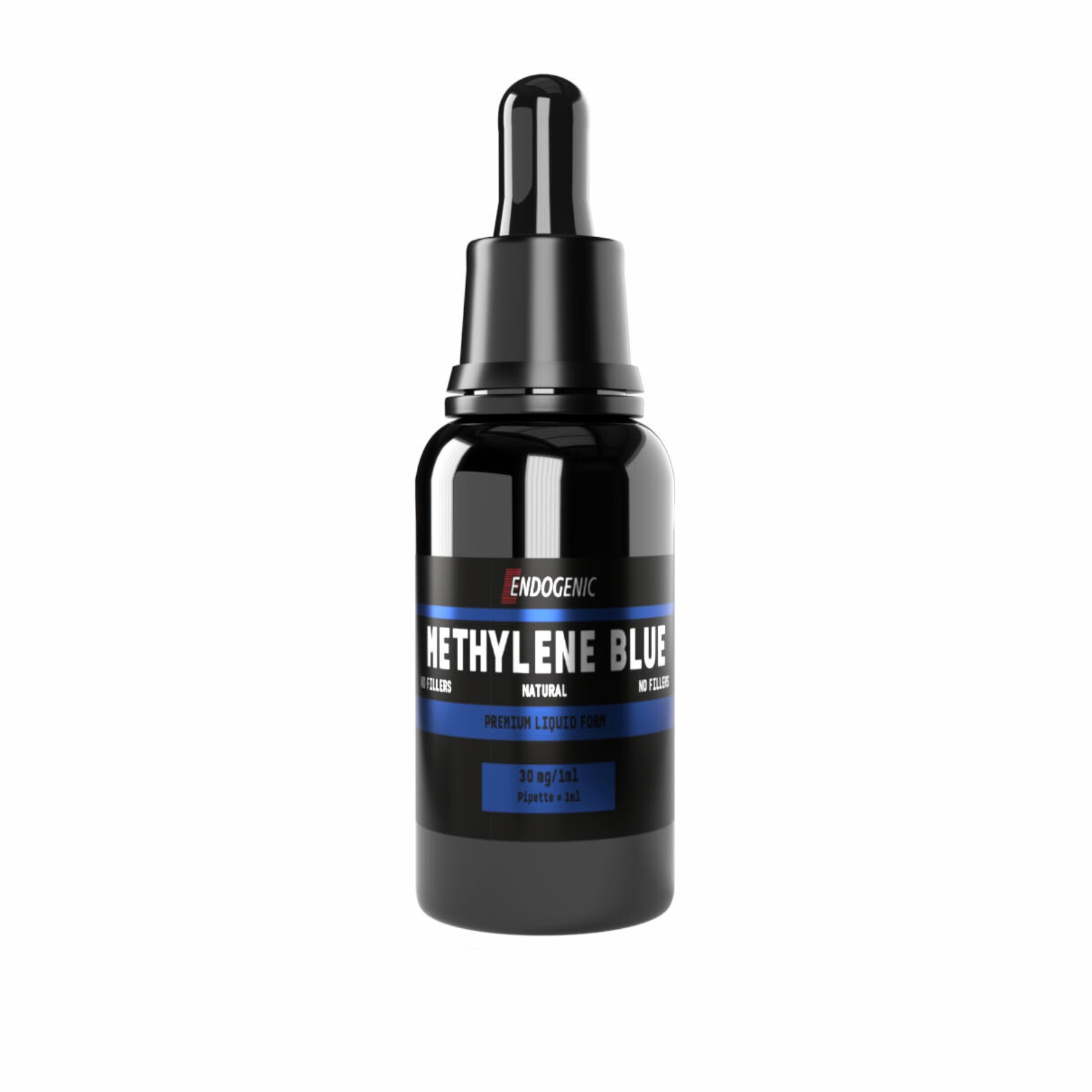
Methylene blue does not have a single receptor. Its profile is the resultant of the redox-active chemistry of a molecule capable of reversibly accepting and donating electrons — and it is precisely this feature that underlies most of the observed effects.
Alternative electron carrier — bypass of complexes I and III
At the heart of methylene blue’s action is the alternating transition between the oxidized form (MB⁺, blue) and the reduced form (leuco-MB, colorless). In the mitochondrion MB⁺ accepts electrons from NADH and from NADPH dehydrogenase, and then donates them directly to cytochrome c — bypassing complex I and complex III of the respiratory chain. In practice: when these complexes are damaged (by toxins, hypoxia, neurodegeneration), methylene blue acts as a makeshift “detour” for electrons, sustaining flow through the chain and ATP production (Rojas et al. 2012). This is the mechanism that explains why the compound attracts interest wherever the problem is a cellular energy deficit.
Increased complex IV (cytochrome c oxidase) activity — and the hormetic curve
At low concentrations methylene blue raises the activity of complex IV (cytochrome c oxidase) — the terminal enzyme of the respiratory chain — increasing oxygen consumption and ATP synthesis in neurons (Tucker et al. 2018). Here the central pharmacological feature appears: an inverted-U-shaped hormetic relationship. Low doses (in rodent models on the order of 0.5–4 mg/kg) support energy metabolism; above this threshold (about 4 mg/kg) the molecule begins to act pro-oxidatively and inhibits mitochondrial respiration.
This is the biochemical justification for the “little, not a lot” rule — escalating the dose not only fails to help, it reverses the effect.
Antioxidant activity
Paradoxically, the same redox activity within an appropriate concentration range allows methylene blue to scavenge reactive oxygen species (ROS) and nitrogen species (RNS) and to reduce excess nitric oxide. The molecule captures electrons from free-radical reactions, limiting oxidative stress in nervous tissue (Schirmer et al. 2011). Together with the bypass of the respiratory chain, this yields a “dual mitochondrial protection” profile: more ATP, less oxidative damage — within a narrow dosing window.
Weak inhibition of MAO-A and MAO-B — the source of the most dangerous interaction
At higher doses methylene blue inhibits monoamine oxidase — the enzyme that breaks down serotonin, noradrenaline and dopamine. Ramsay et al. (2007) demonstrated that it is a potent MAO-A inhibitor with an affinity close to that of classic MAOIs, which makes the interaction with serotonergic drugs a real, not a theoretical, threat. The mechanism is simple and brutal: if MAO is blocked, and at the same time a serotonergic drug increases the concentration of serotonin in the synapse, the serotonin has no way to be broken down — and it accumulates to a toxic level.
This is the biochemical basis of serotonin syndrome and the reason why in 2011 the FDA warned against combining methylthioninium with serotonergic drugs.
IMPORTANT DISTINCTION
“Weak MAO inhibition” does not mean “safe MAO inhibition.” The term “weak” refers to the dose threshold at which the effect becomes significant — not to the strength of the block itself. At the doses used in a nootropic context, the risk of a serotonergic interaction is real. If you are taking any drug that affects serotonin, this single property disqualifies methylene blue from your protocol — regardless of the potential mitochondrial benefits.
Inhibition of tau and β-amyloid aggregation
Methylene blue in vitro inhibits the aggregation of tau protein and of β-amyloid — two markers of the neuropathology of Alzheimer’s disease (Atamna and Kumar 2010). It is precisely this direction that led to the development of the derivative LMTX/hydromethylthionine (leuco-methylthionine), which advanced to phase III clinical trials in Alzheimer’s disease (Wischik et al. 2015). It is worth keeping precise: it was the derivative, not methylene blue itself in the chloride form, that was the subject of large clinical trials — and the results of those trials remain the subject of methodological debate.
Neuroprotection in models of Parkinson’s and Alzheimer’s disease
Tying together the pathways above, in preclinical models of Parkinson’s and Alzheimer’s disease methylene blue showed neuroprotective action: it limited neuronal loss, improved memory parameters in rodents and reduced markers of oxidative stress (Tucker et al. 2018). The mechanism is the sum of effects — mitochondrial energetics plus antioxidant protection plus an effect on the aggregation of pathological proteins. A related neuroprotective profile — though through a different, dopaminergic mechanism — is shown in preclinical studies of Parkinson’s disease by 9-MBC (9-methyl-β-carboline), a β-carboline compound with a BDNF/GDNF neurotrophic profile.